


Nan Wu grew up in a small Chinese city where it was difficult for people to get medical help. Five years ago, her mother received a false-positive for breast cancer. Now, Wu is part of a research effort that could prevent such inaccuracies.
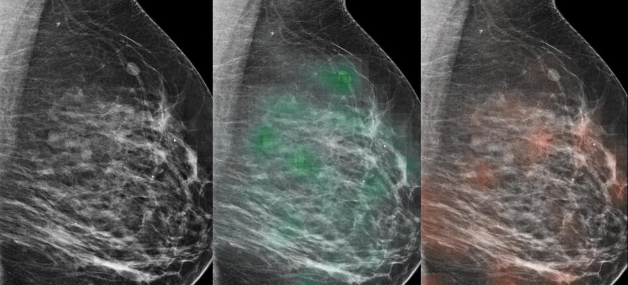
As a second year PhD student and researcher at the NYU Center for Data Science, Wu was involved in a study that developed a new method of detecting the disease, which affects over 200,000 women and results in over 40,000 deaths per year. The method uses artificial intelligence paired with radiologists’ analysis and has a 90% success rate. Researchers from the Center for Data Science and NYU School of Medicine involved hope it will improve early detection, which is essential in preventing deaths.
The AI model used by researchers involves deep neural networks — a technology that mirrors activity of the human brain by using computerized nerve connections. The model also employs machine learning, which allows it to become better at predicting whether or not a patient has cancer as it receives more data.
Although similar to a human’s learning process, NYU School of Medicine Assistant Professor Krzysztof Geras, the study’s senior author, said the AI model is useful because it assesses things in a way radiologists do not, and vice versa.
“A combination of the radiologists with the neural network is actually still better than a radiologist or neural network individually,” Geras, who is affiliated with the NYU Center for Data Science, said. “In practice, radiologists can understand high level structures better while neural networks are better in understanding textural features invisible to the human eye.”
The team trained the AI model using hundreds of thousands of mammograms and images from NYU Langone Health’s 2010-2017 records. The magnitude of data used in the study is significantly more than the norm, as previous research in the field generally used no more than 10,000 images.
“Lots of people worry about why we should use AI in the medical domain,” said Nan Wu, a second year PhD student and researcher at the NYU Center for Data Science. “I think this interestingly shows AI is helping radiologists, not replacing them.”
Dr. Linda Moy is a radiologist at NYU Langone Health who specializes in the detection of breast cancer. She, along with 14 total radiologists, participated in a reader study where the radiologists worked with the AI model and reviewed 720 screening mammogram exams to verify its accuracy.
Moy said the technology could pave the way for increasing doctor’s confidence in breast cancer diagnosis.
“This research is very meaningful to me because we want to find the breast cancer very early before it spreads,” Dr. Moy said. “I’ve had relatives, neighbors, and close friends who’ve been diagnosed with breast cancer. [The model] really helps me more with a decision making process.”
It might take a few years before the technology will assist radiologists regularly across the country and the world, but the team hopes to fine-tune the model to apply it more broadly.
“The model works well with my patients who generally live in Downtown Manhattan,” Moy said. “But it needs to be validated at multiple different locations and on a broad range of patients for us to say it works in a whole variety of settings.”
To encourage this, the team made the model from their paper available online. Since models in the world of machine learning and medical imaging are not usually in the public domain, the team’s decision to share this information is groundbreaking.
“Researchers across the world can download and experiment with them.” Geras said. “We’ve seen hundreds of researchers downloading the model and we are hoping that they will retrain our model for their data or find some creative ways for improving the model.”
Email Roshni Raj at [email protected].